551 results found | searching for "protein,"
-

-
Hey thank you!!! I was seeking for the particular information for long time. Good Luck ? https://forum.gettinglost.ca/user/urgentemiratesvisa https://www.verema.com/usuarios/privado/urgentemiratesvisa https://nootheme.com/forums/users/urgentemiratesvisa/ https://www.amino.dk/members/urgentemiratesvisa/default.aspx https://www.ukwomenorg.com/urgentemiratesvisa https://coraops.com/forums/users/urgentemiratesvisa/ http://blog.boltonvalley.com/2011/12/snow-natural-and-manmade.html?sc=1756201826838#c2893368513953420744 https://momscrazycooking.blogspot.com/2011/09/pioneer-woman-chicken-pot-pie-secret.html?sc=1756202472477#c6061393229208232228 https://nodontpinthat.blogspot.com/2014/06/iodine-and-baby-oil-hair-remover.html?sc=1756202625739#c7405803895343692676 https://sewandthecity.blogspot.com/2013/11/looking-for-cake-pop-stars.html?sc=1756202923315#c53434296083445646 https://juancarikt.blogspot.com/2010/01/realidad-aumentada-el-futuro-en_30.html?sc=1756203018376#c3615995450653782497 https://thecockeyedpessimist.blogspot.com/2019/03/over-cardboard-sea-by-khanh-ha.html?sc=1756203073407#c7870220644783145226 https://divinguniverse.com/user/urgentemiratesvisa https://usbeketrica.com/fr/author/sharma-raguveer https://nothing.community/u/urgentemiratesvisa https://www.mailhilfe.de/author/urgentemiratesvisa https://3rrend.com/urgentemiratesvisa https://www.dmcoffee.com/post/kicking-off-the-nfl-season https://www.sokehsmungovt.org/post/manage-your-blog-from-your-live-site https://gitea.mpc-web.jp/urgentemiratesvisa https://www.biosurfaces.us/single-post/week-21-never-not-working?commentId=20e77dfc-3f86-438d-98fe-ff116fb9b2f4 https://eap.kaspersky.com/user/urgentemiratesvisa https://pad.interhop.org/s/j0-nOPmpD https://www.historiezeme.cz/anglicka-revoluce/#comment-179303 http://www.nlpradio.org/self-improvement/silencing-the-mind/#comment-29325 https://etchrock.com/profile/raguveer-sharma https://mizunosoccershoesfans.com/urgentemiratesvisa https://supplyautonomy.com/urgentemiratesvi.in https://www.abookmarking.com/story/urgent-emirates-visa-get-urgent-dubai-visa-in-1-hour https://www.besport.com/user/924115 https://kakogawa.diycities.jp/profiles/urgentemiratesvisa/activity https://www.theroyalbutler.co.uk/post/christmas-work-party-etiquette-with-next?commentid=c8a251b3-4f6a-4029-b98e-c058a1f04424&commentId=d8b37460-5dc3-45ad-a745-de9e77e3e6d0 https://skincheckchampions.com/ufaq/wheres-the-clinic/#comment-58809 https://www.frenchguycooking.com/pizzadough#comment-117281 https://www.ossklm.si/2014/02/10/slovenski-kulturni-praznik/#comment-47287 https://mediablogstage.prnewswire.com/2022/06/03/media-news-recap-060322/#comment-476665 https://www.overmugged.com/post/how-to-calculate-your-o-levels-l1r5-l1r4?commentId=3d87ad71-1374-4000-9222-da19e0123ebd https://www.waspnation.com/post/w-a-s-p-the-7-savage-boxset-a-7-cd-collection-celebrating-the-explosive-legacy-of-w-a-s-p?commentId=d8efe492-3414-4eb6-81cf-39ad439c1f6c https://www.hillsideautomall.com/2011-hyundai-sonata-4dr-car/#comment-19752 https://choosearecipe.com/butterfinger-balls-recipe-2/comment-page-1351/#comment-102850 https://www.fitlivingeats.com/easy-protein-packed-pasta-salad/#comment-60813 http://somethinghaute.com/beyond-skyscrapers-exploring-dubais-hidden-gems-from-al-seef-to-green-planet/#comment-4333487 https://havingfun.es/doodling/#comment-13917 https://brownbagteacher.com/word-work-sentence-scramble/#comment-166929 https://skincheckchampions.com/ufaq/wheres-the-clinic/#comment-58836 https://dailymoneyout.com/2021/09/14/tips-on-how-to-win-casino-slots-and-baccarat/#comment-535928 https://webrazzi.com/profil/urgentemiratesvisa/ https://www.freebeg.com/forum/member.php?action=profile&uid=119611 https://www.pianobook.co.uk/profile/urgentemiratesvisa/ https://mytravaly.com/blog/blog-read/?t=Prophet+Muhammad+Birthday+UAE+Holiday+2025%3A+Dates%2C+Meaning+%26amp%3B+Travel+Guide https://www.paramedicine.com/post/a-paramedicine-com-experiment-in-crowd-sourced-peer-review-adult-anaphylaxis-ariticle-join-in https://edostate.com/urgentemiratesvisa https://www.crownmaple.com/post/baked-brie-with-crown-maple-organic-maple-syrup-and-pear-confit?commentId=4feafb4d-8337-4452-8219-fdd69564f94e https://www.aloha-poke.com/post/die-sat-1-reportage-berichtet-%C3%BCber-aloha-poke?commentId=1fbc2b76-245c-441d-a672-9f75fb91ed30 http://www.mayricherfullerbe.com/2013/04/vintage-roadrunner-glassware.html?sc=1756372228522#c1339869760356826126 https://www.imcas.com/en/profile/raguveer-sharma https://usedvape.com/profile/raguveer-sharma/ https://tryhackme.com/p/pandaraguveer https://trackrecord.id/urgentemiratesvisa https://yebble.com/profile/raguveer-sharma/ https://www.atrevetesolo.com/blog-de-singles/viaje-de-ensueno-tailandia?page=28#comment-135780 https://manacube.com/members/urgentemiratesvisa.281887/#about https://www.recipesandreviews.co.uk/2013/08/darts-farm-exeter.html?sc=1756378343243#c8940172182166173008 https://provc.gctu.edu.gh/2022/06/13/hello-world/#comment-23623 https://as-cn-video.rockwool.com/by-nature-circular-169#comment115637702 https://www.aussieairwing.com.au/post/aawtrailercomp https://hawksites.newpaltz.edu/jennbrannigan/2015/11/20/hello-world/#comment-61143 https://blogs.umb.edu/jenniferpetruzzi001/2015/11/09/video-essays/#comment-22839 https://www.nomomente.org/post/plant-based-leather?commentId=c99e259f-4f5c-47f7-8459-d4fcc8106e3e https://www.jamesmforrest.co.uk/post/walking-the-national-three-peaks-in-17-days?commentId=bb21792a-afd5-422e-a319-3ee30ad840fa https://www.2chicksandahammer.com/post/family-q-a-with-mina?commentId=4bf0de3c-163a-46bb-9fd5-b7ddd6d4232c https://userstyles.world/user/urgentemiratesvisa https://beforeitsnews.com/v3/contributor/bio/?uid=1024122 https://www.empireslicepizza.com/top-pizza-style-by-state/#comment-133066
-

-
 E. coli Model for Protein Amplification Escherichia coli is a bacterium that most of us know first from recalls of foods like meat and lettuce. Although E. coli is a commensal (non-harmful) gut bacterium, the wrong circumstance and strain can make infection uncomfortable and dangerous. E. coli is gram negative, meaning it has a double-layered cell membrane that makes it more difficult to treat with antibiotics. Our homogenization equipment is regularly used to disrupt E. coli. In “Identification and Biosynthesis of pro-inflammatory sulfonolipids” authors Hou et al. use the NanoGenizer to break apart E. coli after using it for protein expression. Many other researchers have done the same (https://www.genizer.com/u_file/2206/file/6bffa6a456.pdf, https://www.genizer.com/u_file/2312/file/298f8a65e6.pdf , https://www.genizer.com/u_file/2312/file/3ca3c35d8a.pdf) What makes this common cause of diarrheal illnesses a welcome tool in so many research labs? First, we need to understand the research problem E. coli is being used to address in many labs. Researchers often need large amounts of proteins for a variety of purposes. One common example is for pharmaceutical production at a large scale. However, having to obtain that protein from its natural source can be time-consuming, resource intensive and complicated. Instead, researchers will often take the gene that encodes that protein and transform it into a bacterium. There, the bacteria will make that protein more quickly. However, not just any bacteria are suited for this purpose, and that’s what makes E. coli a laboratory star. Speed: E. coli can double as fast as every 20 minutes. Optimal conditions for quickly producing large quantities of protein. Density: E. coli can grow densely, reducing the amount of space required for protein production. Materials: E. coli can grow well on affordable and easily obtainable materials, making it accessible to researchers. Universality: The wide breadth of research done using E. coli as a model makes it easier to work with. Tractability: Tractability refers to the ease of editing a genome. E. coli is naturally genetically tractable and can pick up new genes and add them to itself. This eases the process of adjusting its genome for researchers. As a pathogen, this feature also helps E. coli evade antibiotics. So researchers, go forth and enjoy this model organism in the laboratory, if not always in your kitchen! Sources: https://www.ncbi.nlm.nih.gov/books/NBK564298/ https://pmc.ncbi.nlm.nih.gov/articles/PMC4029002/ https://www.ncbi.nlm.nih.gov/books/NBK562895/
 -
-
Pharmaceutical Giants Rush to Develop PD-(L)1 Bispecific Antibodies: A New Battlefield in Immunotherapy In recent years, immune checkpoint inhibitors (ICIs) have emerged as a significant breakthrough in cancer therapy, reshaping traditional treatment paradigms. PD-1/PD-L1 pathway inhibitors have been widely used across various cancer treatments, demonstrating impressive efficacy. As PD-(L)1 inhibitor therapies continue to mature, pharmaceutical giants have turned their attention to PD-(L)1 bispecific antibodies (BsAbs), a new class of antibody drugs that has become a hot field for development in the industry. The key advantage of PD-(L)1 bispecific antibodies is their ability to target both PD-1 and PD-L1 simultaneously, not only enhancing anti-tumor activity through stronger immune activation effects but also overcoming the limitations of single-target antibodies. As a result, pharmaceutical companies have invested heavily in developing PD-(L)1 bispecific antibodies, striving to achieve breakthroughs in this area. PD-(L)1 bispecific antibodies are one of the brightest stars in antibody drug development. These bispecific antibodies can recognize two different antigens or targets at the same time, resulting in a synergistic effect. In their design, bispecific antibodies not only block the binding between PD-1 and PD-L1 but also recruit immune cells, enhancing the immune system's ability to attack tumors. This "two-pronged" strategy has made PD-(L)1 bispecific antibodies a focal point in cancer immunotherapy. Currently, numerous pharmaceutical and biotechnology companies are actively advancing the clinical research of PD-(L)1 bispecific antibodies, especially in cancer immunotherapy, where they show significant promise. Some PD-(L)1 bispecific antibodies can not only target immune evasion mechanisms within the tumor microenvironment but also significantly improve patient survival, positioning them as the "new favorite" in cancer immunotherapy. The PD-1/PD-L1 Pathway and Mechanism of Immune Escape PD-1 (Programmed Cell Death Protein 1) is a crucial checkpoint in the immune system. By binding to its ligand PD-L1, PD-1 inhibits T-cell activation, regulating immune responses and preventing excessive immune reactions that could harm the body's tissues. However, tumor cells often exploit this mechanism to evade immune surveillance, promoting their growth and metastasis. The role of the PD-1/PD-L1 pathway in immune evasion makes it a key target for immunotherapy. The application of PD-1 and PD-L1 monoclonal antibodies helps to relieve immune suppression, restore T-cell function, and boost the immune system's ability to recognize and eliminate tumor cells. As a result, immune checkpoint inhibitors are widely used in the treatment of various cancers, including non-small cell lung cancer, melanoma, and renal cell carcinoma. Despite the promising clinical efficacy of PD-1/PD-L1 inhibitors, challenges remain. Some patients develop resistance to these therapies, and side effects, such as immune-related adverse events, can complicate clinical application. This has driven researchers and pharmaceutical companies to explore new treatment options, with bispecific antibodies emerging as a promising solution. In the development of immunotherapy drugs, the use of cell models plays a critical role. Human PD-1 recombinant cell lines are among the most essential tools for studying the PD-1 pathway, widely used for drug screening, mechanistic research, and preclinical evaluation. By stably expressing the PD-1 protein in cells, researchers can simulate interactions between immune cells and tumor cells, explore the mechanisms of the PD-1/PD-L1 pathway, and evaluate the efficacy of PD-1/PD-L1 targeted therapies. For example, using these recombinant cell lines, researchers can simulate immune escape processes in the tumor microenvironment, investigate the mechanisms of action of PD-1 inhibitors, and screen new antibody drugs. This tool is also crucial in evaluating the preclinical potential of drugs, contributing to the advancement of anti-tumor immunotherapies. As the field of immunotherapy continues to evolve, the clinical application of PD-1/PD-L1 inhibitors has made significant strides. However, several challenges remain, particularly related to individual variation, resistance, and side effects. Researchers are actively exploring combination therapies to enhance treatment outcomes, such as combining PD-1 inhibitors with chemotherapy, targeted therapies, or vaccines. This may help overcome resistance and improve the overall efficacy of treatment. Furthermore, as new immunotherapy strategies emerge, the application of PD-1 and related treatments may extend beyond cancer. Immune checkpoint inhibitors are showing promise in autoimmune diseases, infectious diseases, and other areas, making them a key focus in future medical research. From the early days of single-target therapies to the current focus on bispecific antibodies, immunotherapy continues to innovate, transforming cancer treatment approaches. With the emergence of PD-1 recombinant cell lines and new immunotherapy solutions, we can look forward to a new era in cancer therapy, where more patients will benefit and the full potential of immunotherapy will be unlocked. https://www.creative-biolabs.com/immuno-oncology/human-pd-1-recombinant-cell-line-jurkat-2696.htm
-
-
New Insights Into the Pathogenesis and Diagnosis of Rheumatoid Arthritis The hallmark of rheumatoid arthritis (RA) is erosive arthritis, an autoimmune disease that ultimately results in joint deformities and functional loss. It can also be complicated by pulmonary disease, cardiovascular disease, malignant tumors, and depression. The etiology of RA remains unclear. However, infections have been suggested as environmental triggers in as many as 20% of patients. Due to its perplexing etiology, a more detailed exploration of the pathogenesis of RA has been presented in an article titled "Altered antibody response to Epstein-Barr virus in patients with rheumatoid arthritis and healthy subjects predisposed to the disease" published in Immunol. The article delves deeper into the potential connection between Epstein-Barr virus (EBV) and RA, employing dependable tests that quantify antibodies directed against specific EBV antigens. So why did the research team link EBV to the development of RA? A disease similar to RA called polyarticular arthritis is induced by various viral infections, including rubella, HTLV-1, parvovirus B19, etc. Given that EBV has been connected with other autoimmune diseases such as multiple sclerosis and systemic lupus erythematosus, it is reasonable to assume that this virus may also be related to the pathogenesis of RA. Therefore, this article investigates the EBV antibody patterns in rheumatoid arthritis patients to assess the heritability of the antibody responses to the EBV-encoded EBNA1 protein, ultimately concluding that the levels of EBNA1 antibodies are notably dissimilar in RA patients compared to healthy individuals. Nevertheless, the findings reached in this article represent just a fraction of the complex investigation into the etiology of RA. Undoubtedly, the uncertain underlying causes of RA pose challenges for accurate diagnosis. RA can affect individuals of any age, but it is most frequently diagnosed in individuals between the ages of 35 and 50. Early diagnosis of RA can help identify people at risk of RA and prevent complications and disease progression. Modern imaging techniques, such as X-rays, magnetic resonance imaging, and ultrasound, aid in diagnosing RA by capturing images of affected joints. However, these methods are challenging for early RA diagnosis due to the similarity of early symptoms with those of other diseases. Additionally, detection methods that use serum markers, such as the anti-cyclic citrullinated peptide test in combination with rheumatoid factor, can improve the final diagnosis of patients with negative results from routine tests. As an efficient and precise method, IVD immunological assays and test kits rely on the specific recognition between one or more antibodies and an antigen, allowing for the detection and quantification of various antibodies in different types of samples (including serum, urine, saliva, environmental media, and more). Specifically, some rheumatoid arthritis biomarkers that have been developed for early diagnosis of RA include but are not limited to UH-RA 1, UH-RA 9, UH-RA 14, UH-RA 21, Rheumatoid Factor, 14-3-3 Eta Protein, PAD4, etc. Not only are RA biomarkers evolving, but so are their development solutions in the following approaches: * IVD Antibody Development * Antibody Pair Development * Antibody & Protein Conjugation * IVD Immunoassay Development https://www.creative-biolabs.com/drug-discovery/diagnostics/biomarker-and-antibody-development-for-rheumatoid-arthritis.htm
-
-
Decoding Cellular Signals: The Power of Phosphorylation Antibody Arrays in Modern Biology Inside every cell, complex communication networks are constantly at work. These systems—known as signaling pathways—allow cells to respond to changes in their environment, control growth, defend against threats, and carry out essential biological tasks. One of the key methods cells use to transmit signals is phosphorylation, a process where a phosphate group is added to a protein to change its activity. Phosphorylation acts like a molecular switch. When certain proteins are phosphorylated, they may become active, move to a new part of the cell, or interact differently with other molecules. Because this process is so vital to healthy cell function, it's no surprise that disruptions in phosphorylation can lead to diseases such as cancer, diabetes, and autoimmune disorders. To understand these changes, researchers turn to phosphorylation antibody arrays, which allow them to track the activation of many signaling proteins in one simple experiment. Understanding Insulin Signaling with Antibody Arrays One major pathway that scientists often study is the insulin receptor signaling pathway, which controls how cells take in and use glucose. When this system works properly, cells respond efficiently to insulin. But when something goes wrong, it can lead to insulin resistance or type 2 diabetes. The Human Insulin Receptor Pathway Phosphorylation Antibody Array is specially designed to measure the phosphorylation levels of key proteins in this pathway. With this array, researchers can monitor how well the insulin signal is transmitted within the cell—information that is vital for diabetes research and drug development. Tracking Cell Survival Signals in the AKT Pathway Another pathway closely tied to cell growth and survival is the AKT signaling pathway. This pathway, also called the PI3K/AKT pathway, is often overactive in cancer cells, allowing them to avoid normal controls like apoptosis (programmed cell death) and continue dividing unchecked. The Human AKT Pathway Phosphorylation Antibody Array allows researchers to assess the phosphorylation status of multiple AKT-related proteins. By using this array, scientists can see how strongly the pathway is activated, how it responds to external factors, and how it might be affected by drugs targeting cancer cells. Investigating Immune Responses Through NFκB Signaling Beyond metabolism and cell survival, many researchers focus on inflammation and immune responses. One of the most critical pathways in this area is the NFκB (nuclear factor kappa-light-chain-enhancer of activated B cells) pathway. It helps regulate the body's defense mechanisms, but when dysregulated, it can lead to chronic inflammation or autoimmune disease. The Human NFκB Pathway Phosphorylation Antibody Array is a valuable tool for studying how this pathway behaves under different conditions. It captures a range of phosphorylated proteins involved in the activation and regulation of NFκB, offering insights into inflammation-related diseases and potential treatments. Shared Advantages Across All Three Arrays Even though these arrays target different pathways, they share several key features: Phospho-specific detection: They only detect proteins when they are phosphorylated, giving researchers a real-time picture of pathway activation. High-throughput format: Instead of analyzing one protein at a time, these arrays allow for the simultaneous detection of dozens of phosphorylation events, saving time and providing a broader understanding of cell signaling. User-friendly design: These arrays are ready-to-use with standardized protocols, making them accessible even for labs that don't specialize in proteomics. From Lab to Life: Why It Matters Understanding how cellular signals work — and how they malfunction — is at the core of modern biology and medicine. Phosphorylation antibody arrays make this process more accessible and informative. Whether studying insulin resistance in diabetes, cell survival in cancer, or inflammation in autoimmune diseases, these arrays provide researchers with a powerful window into the signaling activity inside our cells. As we continue to explore the inner workings of the human body, tools like these will be essential for discovering new therapies, personalizing treatments, and advancing precision medicine. https://www.antibody-creativebiolabs.com/akt-pathway-phosphorylation-antibody-array-630290.htm
-
-
Single-Cell CyTOF and Multi-Omics: Decoding the Complexity of Life One Cell at a Time In recent years, single-cell analysis has emerged as a powerful approach to dissect the biological heterogeneity that exists even within a seemingly uniform population of cells. Two cutting-edge technologies—single-cell mass cytometry (CyTOF) and single-cell multi-omics—are leading the way in helping researchers understand how cells function, interact, and change over time in development, disease, and therapy response. What Is Single-Cell CyTOF? Single-cell mass cytometry, or CyTOF, is a hybrid technology that combines the strengths of flow cytometry and mass spectrometry. Instead of using traditional fluorescent tags, CyTOF labels antibodies with heavy metal isotopes, allowing simultaneous measurement of over 40 markers per cell without spectral overlap. This means researchers can obtain highly multiplexed data from millions of cells—ideal for deep immune profiling, stem cell research, or monitoring disease progression. Because each antibody is conjugated to a unique metal tag, the readout is not affected by autofluorescence or signal spillover. This results in much clearer, more accurate data, especially when studying complex systems like the tumor microenvironment or autoimmune conditions where diverse cell types coexist in dynamic states. Going Beyond Proteins: Enter Single-Cell Multi-Omics While CyTOF is ideal for studying the protein landscape of a cell, single-cell multi-omics dives even deeper by integrating multiple layers of cellular information—such as DNA (genomics), RNA (transcriptomics), chromatin accessibility (epigenomics), and proteins (proteomics). By capturing two or more of these data types from the same individual cell, multi-omics techniques offer a more comprehensive understanding of gene regulation, lineage commitment, and cellular state. For instance, combining scRNA-seq (single-cell transcriptome sequencing) with ATAC-seq (assay for transposase-accessible chromatin) can not only reveal which genes are being expressed, but also explain why they are active, based on the accessibility of their promoter and enhancer regions. Such insight is essential when studying processes like cancer metastasis or immune exhaustion. Applications in Research and Medicine Single-cell CyTOF has already made a major impact in immunology. By profiling the expression of surface and intracellular proteins, scientists can classify immune cell subsets, monitor activation states, and track changes in response to infection or immunotherapy. For example, CyTOF has been widely used to study immune responses to COVID-19 vaccines and to characterize T-cell exhaustion in chronic viral infections and tumors. Multi-omics, on the other hand, is particularly powerful for studying developmental biology, neurodegeneration, and epigenetic disorders. In cancer research, it can help identify tumor subclones with distinct regulatory features that might respond differently to treatment. In regenerative medicine, multi-omics can reveal the transcriptional and epigenetic dynamics guiding stem cell differentiation. Integration for Deeper Insights The real magic happens when CyTOF and multi-omics approaches are integrated. By aligning high-dimensional protein expression data with transcriptomic and epigenetic profiles, researchers can build detailed models of cellular behavior and interactions. This is especially valuable in tumor biology, where immune cells, stromal cells, and malignant cells engage in complex cross-talk. For instance, using CyTOF to identify exhausted T-cell phenotypes and multi-omics to characterize their epigenetic signatures can help pinpoint targets for reactivation, guiding the development of next-generation immunotherapies. Final Thoughts As biology becomes increasingly data-rich, the need for high-resolution, multi-dimensional tools continues to grow. Single-cell CyTOF and multi-omics are not just technologies—they’re windows into the hidden lives of cells. Together, they are unlocking the secrets of development, immunity, and disease, one cell at a time. https://singlecell.creative-biolabs.com/single-cell-mass-cytometry-cytof.htm
-
-
 Complement testing evaluates the immune system’s functionality by analyzing pathways and protein activity. It includes assays for classical, lectin, and alternative pathways, as well as detection of activation products and genetic variants. These tests support research in autoimmune disorders, inflammation, and therapeutic development by offering insights into complement regulation, deficiencies, and inhibitor efficacy across diverse biological contexts.https://www.creative-biolabs.com/complement-therapeutics/complement-testing-services.htm
 -

-
 Healthy eating emphasizes a balanced diet rich in nutrient-dense foods, including fruits, vegetables, whole grains, and lean protein sources. It also involves limiting added sugars, saturated and trans fats, and sodium. https://downshiftology.com/30-healthy-recipes-30-minutes-or-less/
 -

-
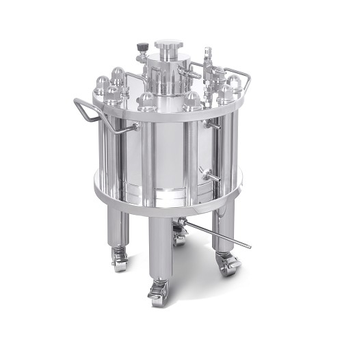 Genizer Jacketed Liposome Extruders for GJE-10000mL The Genizer Jacketed Liposome Extruders is compressed air-driven and temperature-controlled, from testing to manufacturing production of liposome and lipopolyplex, for drug and gene delivery. Homogeneous samples can be produced quickly and easily in minutes; typically 5-10 passes are sufficient to produce a sample with uniform liposome size. Genizer Jacketed Liposome Extruders are designed to generate homogeneous unilamellar liposomes and Lipopolyplex. Extrusion is a one-step procedure that produces liposomes by forcing lipid suspensions of drug, protein or gene through track-etched filters of defined pore size. The process is relatively gentle, exposing the formulation to moderate pressures up to 500 psi at controlled temperatures. The Genizer Jacketed Extruder offers virtually no dead-volume, is easy to clean, suitable for sterilization and in compliance with FDA and GMP sanitary standards in pharmaceuticals. Application: Liposome extrusion Performance: No dead volume The Jacketed Extruder can control the temperature of the extrusion https://www.genizer.com/jacketed-liposome-extruder_p0005.html
 -
-
Collagen Peptide Market and The Applications The collagen peptide market involves small amino acid chains. These are sourced from collagens which are a protein found in the skin or bones. The process involves breaking down the complete components into tinier fragments that are easy for our body to absorb. Fore more info:- https://plaza.rakuten.co.jp/mubazray/diary/202502260000/
Copyright 2019 © P-tweets Refunds